12 April 2024
John Persinger Brings a Wealth of Radiology Experience to the Photomedicine Research Program
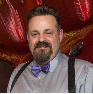
John Persinger, BA, RDMS, RMSKS, CTT+, has more than 35 years of experience in radiology and imaging. He enlisted in the US Army in 1985, attended ultrasound school a few years later, and worked as a staff sonographer in community hospitals and imaging centers for almost 10 years before taking a position as a radiology ultrasound supervisor at Madigan Army Medical Center, which he held for seven years. He has been with Geneva as a musculoskeletal (MSK) Sonographer in the Photomedicine Research Program (PBM) since 2019. In 2022, he opened his own business that focuses on training and mentoring others in MSK ultrasound. Last year, John published the first in a series of handbooks about performing MSK ultrasounds titled “Musculoskeletal Ultrasound Foundations Series Volume I: Shoulder Ultrasound.”
We spoke to John about his experience in radiology and its impact on military medicine.
Describe your role with Geneva and your impact on the program you work on.
I am a musculoskeletal sonographer with The Geneva Foundation, which is an imaging specialist in ultrasound with an additional registry in MSK. This role allows me to draw upon the knowledge and experience I’ve gleaned from 37 years of clinical diagnostic imaging to assist in the selection and set-up of equipment, train staff members in newer ultrasound applications, and conduct ultrasounds. For the PBM program, I worked to ensure we obtained an ultrasound system capable of performing the types of imaging applications that were clinically relevant, oversaw the acquisition and set-up of a cloud-based system through which studies can be stored, shared, or viewed remotely in real-time, and utilized system-based technology to create an image checklist based on the study protocol to standardize and streamline imaging across all study members conducting ultrasounds.
What projects, initiatives, or conferences are up next for you?
During my time here with Geneva, I was involved with several studies utilizing ultrasound at Walter Reed. I am currently engaged with the PBM 08 (plantar fasciitis), PBM 10 (Achilles tendinopathy), and PBM 17 (elastography and microvascular flow normative data) projects being conducted at Madigan.
In April 2024, Dr. Nelson Hager, Dr. Geoff Gabler, and I will attend the annual meeting of the American Institute of Ultrasound in Medicine (AIUM) to support four abstracts from the PBM team. In preparation for the Military Health System Research Symposium (MHSRS) 2024, I am coordinating the abstract submission for PBM 17, which discusses the normative data for elastography and microvascular flow in the gastroc-soleus complex.
What recent work can you share that you are most proud of?
It’s hard to narrow this down to one, as I am proud of many aspects of this role.
On a personal level, to help meet our numbers in terms of participants, I reached out to an MSK radiologist I used to work with at Madigan about this project. We had already sought him out for another PBM project, and he joined as an AI. He brings another clinical skillset to the team, expanding the depth of the recruiting into Radiology and broadening our audience when the findings are published. Expanding the team and seeing them learn from one another is rewarding.
The project is now underway, and when I see each of the scanning members of the team seamlessly working through the steps to complete the study, I know my presence here made a difference in the team’s ability to obtain data to make a difference clinically.
What imaging technology are you currently working with?
I am currently working on two aspects of ultrasound imaging that may prove helpful in diagnosing and assessing therapies for musculoskeletal conditions.
The first is microvascular flow (MVF), an advanced form of color Doppler used to detect the presence of blood flow by assessing the motion of the red blood cells, much like Doppler radar looks for water particles in the atmosphere. We are using MVF to detect neovascularization in tendinopathy.
The other technology is elastography, which uses an ultrasound to determine the relative stiffness of a tissue by pushing a pulse and then measuring the speed of the waves generated in the tissue—like ripples in a pond when you toss in a rock. In a previous role, I taught people how to use this technology in the liver. To see it now expand its potential into MSK is exciting to be part of, especially given the volume of participants we are evaluating. I can see the use of elastography expanding and adapting to other tissues based on what we are doing.
Disclaimer: The views expressed do not reflect the official policy of the Army, the Department of Defense, or the U.S. Government.

I know my presence here made a difference in the team’s ability to obtain data to make a difference clinically.
John Persinger, BA, RDMS, RMSKS, CTT+


